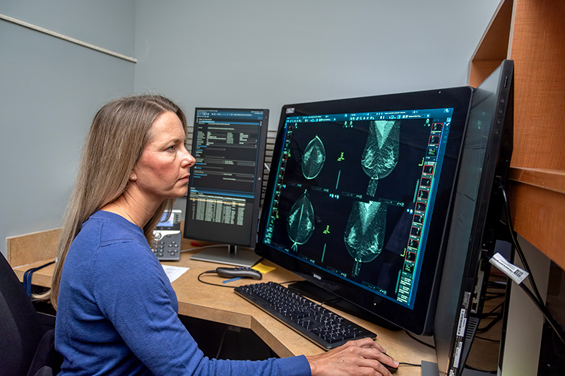
For decades, mammograms have relied on one essential element – the trained eyes of a radiologist searching for tiny abnormalities hidden within shades of black, white and gray.
Now, those radiologists have a new partner.
Artificial intelligence is rapidly becoming an essential tool in breast imaging. In hospitals and imaging centers across the country, AI-enhanced mammography systems help physicians detect cancers earlier, reduce false alarms, and identify subtle warning signs that even experienced readers might miss.
“What we are looking for in breast imaging is improved cancer detection and a decrease in false-positive rates,” said Cleveland Clinic Indian River Hospital radiologist Dr. Cimmie Shahan. “All of that has been constantly improving over the last couple of decades. We went from screen mammography to digital mammography to 3D mammography, and we’ve seen advances in other imaging modalities such as ultrasound and breast MRI. Not only is the image quality better, but our ability to interpret the images is improving as well.”
Experts emphasize that today’s AI systems are not replacing radiologists. Instead, they function as a highly sophisticated second set of eyes, analyzing mammograms with remarkable speed and consistency.
“Radiologists reading mammograms and other breast imaging studies are subspecialty trained or read a very high volume of breast imaging,” she said. “AI algorithms are used like a second reader rather than replacing a trained radiologist.”
Modern AI programs are trained by scanning hundreds of thousands of mammographic images.
By comparing patterns found in previous cancer cases with new scans, the software can identify suspicious areas that require closer attention. However, a human radiologist is still needed to interpret the findings and compare them with a patient’s prior imaging studies.
Multiple studies have shown that AI-assisted mammography can improve cancer detection while sometimes reducing false-positive findings. Research from large screening programs in Europe found that AI helped identify more cancers, including clinically significant tumors, without substantially increasing unnecessary callbacks for patients.
The reason is simple – AI excels at recognizing patterns.
A radiologist may notice a suspicious cluster of calcifications or a subtle distortion in breast tissue. AI can analyze the same image pixel by pixel, comparing it against enormous databases of previous exams and identifying abnormalities that may be too subtle for the human eye to easily recognize.
Researchers have found that AI and radiologists often detect different warning signs, making the combination of both more powerful than either working alone.
This advantage may be especially important for women with dense breast tissue.
Dense breasts contain more glandular and fibrous tissue and less fat, making cancers harder to detect because both dense tissue and tumors appear white on a mammogram. AI systems can help highlight subtle areas of concern hidden within this complex tissue, potentially improving detection for women who have historically been among the most challenging to screen.
Beyond simply finding cancer, AI is beginning to do something even more remarkable – predict risk.
Researchers are developing systems capable of estimating a woman’s likelihood of developing breast cancer years before any tumor is visible. By analyzing subtle imaging characteristics that humans cannot readily interpret, these programs may eventually allow screening schedules to be tailored to individual risk rather than relying on a one-size-fits-all age and family-history indicated approach.
Imagine two women of the same age. One may have imaging features suggesting a very low risk of developing cancer over the next several years, while another may show patterns associated with significantly higher risk. Future AI systems could help determine who needs more frequent screening, supplemental MRI scans or closer surveillance.
“What the AI algorithm is determining is a pattern that would be typical of, or more suggestive of, breast cancer,” Dr. Shahan explained. “It might recognize the distribution or morphology of calcifications that are more suspicious than others or identify a distorted mass. But if a patient has a history of trauma or surgery to the breast, it could create the exact same appearance.
Human radiologists are able to recognize and incorporate the patient’s history and understand that it isn’t truly a suspicious abnormality.”
That distinction highlights why human expertise remains indispensable.
Radiologists do far more than identify abnormalities on images. They consider patient history, compare prior studies, correlate findings with ultrasound and MRI examinations, communicate with referring physicians, and help guide biopsies and treatment decisions. Human judgment is essential when imaging findings are ambiguous or when multiple clinical factors must be considered.
Current AI systems also have other limitations. They can miss unusual cancers or perform less effectively in populations that differ from the data on which they were trained. Regulatory agencies and medical organizations continue to stress that human oversight is necessary. Most experts envision AI serving as an assistant rather than an autonomous decision-maker.
Even so, AI could eventually help triage screening exams by rapidly identifying normal studies and directing radiologists’ attention to higher-risk cases. This could help address a growing shortage of breast imaging specialists while reducing delays in reporting results.
“It has become more difficult for breast imaging practices to keep up with the volume,” Dr. Shahan said. “That is definitely a feature of artificial intelligence that is attractive to many practices because it can help improve workflow efficiency.”
The most important takeaway is that medicine is gaining a powerful new tool, not replacing the professionals who use it.
Breast cancer remains one of the most common cancers affecting women worldwide, and early detection continues to be one of the most important factors in improving survival. If artificial intelligence can help find cancers sooner, reduce missed diagnoses, and provide radiologists with greater confidence in their interpretations, it may become one of the most significant advances in breast imaging since the introduction of digital mammography.
Dr. Cimmie Shahan attended George Washington University School of Medicine in Washington, D.C. and completed a residency in diagnostic radiology and a fellowship in breast imaging at West Virginia University. She is one of four breast imaging specialists with the radiology department at Cleveland Clinic Indian River Hospital and Vero Radiology. For radiology appointments, call 772-562-0163.
Photos by Joshua Kodis