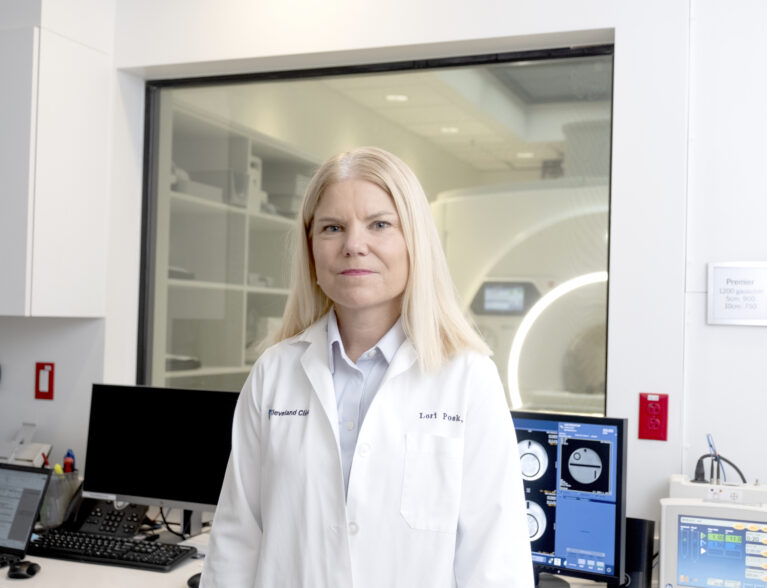
Cleveland Clinic Indian River Hospital has upgraded its magnetic resonance imaging (MRI) scanners to offer academic medical center-level imaging for diagnosis and management of a wide range of medical conditions.
“We have spent the last few years upgrading the imaging capability of radiology, both in terms of the quality of the scans and the complexity of what we have to offer to patients and the community in Vero Beach; both through new protocols, meaning the type of imaging details, how we scan the cases, all the way through to the hardware and the software that we have available to produce and process them,” said Dr. Avi Oppenheimer, a neuroradiologist with Cleveland Clinic.
“These upgrades enable my clinical colleagues to make more precise diagnoses and to treat and monitor their patients in a better way than in the past.”
Vero Radiology Associates, which is part of Cleveland Clinic, now has a 3T MRI scanner with a GE Premier magnet. MRI magnets are measured in their strength of the magnetic field, which is measured in Tesla, and the 3Tesla MRI is one of the strongest magnets available for imaging.
This scanner produces a magnetic field twice as strong as that of a standard 1.5 MRI scanner.
“This new software allows the scanner to scan faster, so the patient is in the scanner for less time,” Dr. Oppenheimer explained. “The AI technology allows it to scan with a high resolution and more detail than other scanners so we’re able to get better quality images faster.”
According to the National Cancer Institute, the 3T MRI uses radio waves and a strong magnet linked to a computer to create detailed images of areas inside of the body. These pictures can show the difference between normal and abnormal tissue. It is used to make images of the brain, the spine, the soft tissue of the joints, and the inside of bones and blood vessels. The 3T MRI is beneficial in the diagnosis of neurological conditions such as multiple sclerosis, brain tumors, Parkinson disease, epilepsy, traumatic brain injuries, and Alzheimer’s and other forms of dementia.
The stronger magnetic field of a 3T MRI allows for clearer more precise images, which is particularly useful for visualizing small anatomical structures, subtle abnormalities or lesions that may not be visible with a lower-field MRI scanner, making it an invaluable tool for assessing soft tissue and spinal injuries, liver and pancreatic conditions, pelvic disorders and cancers of the breast and prostate.
Dr. Oppenheimer explained how the 3T MRI has revolutionized the way neurologists diagnose memory loss diseases.
“Dementia is a very common issue, but it’s difficult to diagnose,” he said. “Radiologists generally use a qualitative assessment and standard imaging to get a sense of brain atrophy and brain volume loss. We’re now able to scan the brain with a 3D technique that images the brain as a volume as opposed to individual thicker slices. That detail and the 3D technique allows that data to be processed by a software program called NeuroQuant, which segments the brain using a computer algorithm into different substructures and then compares the patient’s volume of their brain to a standard age matched population. We know that the brain does shrink with time, but the question is, has this patient’s brain shrunk more than their age cohort?”
This advanced imaging technique creates a way to track a patient’s brain volume and see how it changes in volume over time. Comparing the scans from year to year can track the amount of atrophy and where the atrophy occurs. A very detailed report allows clinicians to see exactly what is taking place and how it compares to the patient’s age group.
“Clinically, one of the areas that we look at, particularly with memory loss and Alzheimer’s disease, is an area of the brain called the hippocampus,” added Dr. Lori Posk, a primary care physician with Cleveland Clinic Indian River Hospital. “The hippocampus is a small part of the brain but it does a very big job with learning and memory. We will often see more shrinkage of the hippocampus in patients with Alzheimer’s disease, so this is one potential biomarker we can follow over time.”
Dr. Posk typically orders an MRI for any patient concerned with memory loss.
“This helps us rule out other causes of memory loss that are rare, such as brain tumor or subdural hematoma, but with this new software we can pinpoint the atrophy to a certain area and correlate that to age-matched controls and look at that over time. This can help in the evaluation of a patient that we suspect might have Alzheimer’s disease.
“Typically, we see the patient, examine them, order some cognitive testing and possibly some bloodwork, and then often order the MRI based on those findings to help in the diagnosis of memory loss. Now we have the option of using this NeuroQuant software to compare the brain volumes to those of standard patient population. It is used in conjunction with other tools when I’m looking at making the diagnosis of dementia or Alzheimer’s disease.”
A complete software upgrade to the hospital’s 1.5T MRI scanner provides access to the type of imagining protocols found in major academic medical centers.
“We’ve also upgraded the hospital’s 1.5T MRI scanner and implemented very high-resolution imaging of the skull base, which was previously not available in Vero Beach,” Dr. Oppenheimer said. “We use this imaging to see the cranial nerves such as the trigeminal nerve and the facial nerve. Those nerves are about a millimeter in size, or about the size of dental floss, so the imagining has to be on that level to be able to see the nerve and the pathology associated with it to make a diagnosis.
“The new imagining is imaging the skull base that is sub-millimeter in slice thickness so we are looking at images that are somewhere between 600-800 microns in thickness, almost like what a microscope would do. This is very helpful for treating patients with trigeminal neuralgia and identifying different causes of that disorder or facial nerve palsy (Bell’s Palsy) and other neurological disorders.”
Dr. Oppenheimer and Dr. Posk both stress that the upgraded technology offers a level of imaging to Vero patients who might have had to travel out of the area for such advanced imaging in the past.
Dr. Avi Oppenheimer is a neuroradiologist based in Cleveland Clinic’s Weston Hospital who interprets radiology reports for Cleveland Clinic hospitals. He received his medical degree from Albert Einstein College of Medicine and completed a residency in Internal Medicine at Staten Island University Hospital and in diagnostic radiology at Montefiore Medical Center in Bronx, N.Y. His fellowship in Neuroradiology was completed at Johns Hopkins Hospital.
Dr. Lori Posk earned her medical degree from Michigan State University in East Lansing and completed her internship and residency in Internal medicine at Cleveland Clinic in Cleveland. Her office is located in the Rosner Family Health and Wellness Center, 3450 11th Court, Vero Beach. Call 772-794-3364 for an appointment.


