
A strange sounding eye disease – Fuchs’ dystrophy – is not well known despite being fairly widespread, affecting as many as 4 percent of people over the age of 40.
People with Fuchs’ dystrophy often wake up in the morning with distorted vision, like looking through a steamy shower door. It’s blurry, hazy and frustratingly unclear. This progressive eye disease quietly affects the cornea’s inner workings, often showing no signs until it disrupts daily life.
Also known as Fuchs’ endothelial corneal dystrophy, the disease is a hereditary eye condition that causes fluid buildup in the cornea, leading to vision problems like blurred vision and sensitivity to light.
“Fuchs’ dystrophy is a progressive condition that targets the innermost layer of the cornea known as the endothelium,” said Dr. Christopher Seery of New Vision Eye Center. “The endothelium is responsible for maintaining fluid balance in the corneal stroma – the thickest and most important layer of the cornea. When endothelial cells fail, it leads to corneal edema, resulting in hazy vision, light sensitivity, and decreased visual contrast.
“To simplify, think of the eye like a camera,” Dr. Seery continued. “There’s a lens and a focusing element – essentially the cornea. If anything interferes with that focusing process, such as cell damage or fluid buildup, light scatters, and the result is blurred vision or sensitivity to light.”
According to the Corneal Dystrophy Foundation, Fuchs typically emerges in middle age, although symptoms may not appear right away. It’s caused by the gradual loss of “pumper” cells that keep fluid from accumulating in the cornea. Without these cells, fluid builds up, swelling the cornea and distorting vision – especially in the morning. Over time, tiny blisters can form and burst, causing significant discomfort.
Patients often describe a gritty or sandy sensation, as if something is stuck in their eye. They also experience glare, halos and increasing light sensitivity. As the disease progresses, daily fluctuations in vision stop, and significant vision loss may occur. In advanced cases, a corneal transplant may be necessary.
“There are different types,” Dr. Seery said. “Some are aggressive and show up in a person’s 30s or 40s, but most develop slowly and aren’t diagnosed until after age 50. Surgery – particularly cataract surgery – can sometimes trigger or worsen the condition. That’s why we weigh all surgical options carefully when treating someone with advanced Fuchs and will sometimes combine the cataract surgery with the cornea surgery.”
Diagnosis involves several specialized tests. Eye doctors use a slit lamp to spot tiny bumps called guttae on the back surface of the cornea. They’ll also assess swelling and the stage of the disease. Corneal pachymetry measures the thickness of the cornea, while tomography provides detailed images to help track changes over time.
“A clinical exam is still the gold standard for early diagnosis,” urges Dr. Seery. “I recommend regular eye exams beginning at age 40 – even if you have perfect vision – because early detection allows us to be proactive rather than reactive.”
Initial treatment often starts with hypertonic saline drops or ointments, which draw fluid out of the cornea. However, if these prove ineffective, surgery becomes the next step.
“The surgical options are evolving quickly,” Dr. Seery said. “The gold standard is endothelial keratoplasty (EK), which is less risky than a full-thickness transplant.” He explained that EK comes in two forms: DSEK (Descemet’s Stripping Endothelial Keratoplasty) and DMEK (Descemet Membrane Endothelial Keratoplasty). DMEK uses a thinner, more delicate layer for transplant, while DSEK includes a bit of extra tissue for added stability.
“Untreated Fuchs can turn into a debilitating vision loss,” Dr. Seery continued. “But the good thing about the cornea is that, just like the lens on the camera, it can be exchanged. When a cornea is so swollen that it is truly hazed over, it can develop scarring. That’s when the full thickness transplant becomes necessary. But as long as there’s just swelling with minimal scar formation, then one of the endothelial transplants can usually be done.”
One newer surgical approach, called DSO (Descemet Stripping Only), involves removing only the damaged cells without replacing them and allowing the healthy cells to compensate for the removed areas.
“We can actually just remove the damaged cells with no transplant and then that natural cell population from the rest of the cornea can migrate and eventually improve vision,” Dr. Seery explained. “By removing the tissue in a localized area, the peripheral cornea can be preserved and the normal cells in the periphery can compensate for the removed areas. Just removing them can be near curative.”
Emerging treatments include experimental drugs aimed at regenerating endothelial cells and cell-based injections that may one day offer a non-surgical option for managing the disease.
Fuchs’ dystrophy may blur the world around you but with early detection and rapidly advancing treatment options, clearer days are in sight.
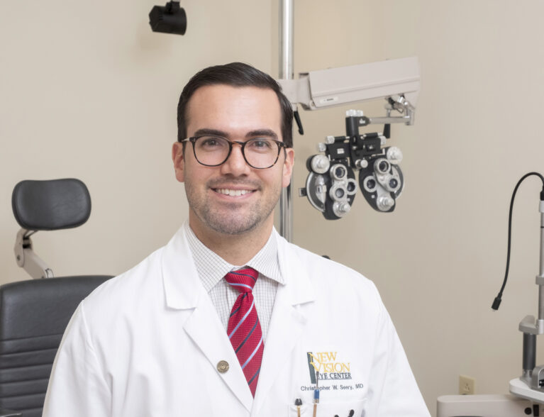
Dr. Christopher Seery earned his medical degree and completed his internship and ophthalmology residency at Rutgers New Jersey Medical School. In order to gain the expertise as a cornea and refractive surgeon, he completed his fellowship in cornea, external disease and refractive surgery at the prestigious Bascom Palmer Eye Institute at the University of Miami. He is accepting new patients at New Vision Eye Center, 1055 37th Place in Vero Beach. The phone number is 772-257-8700.