Just like the windshield of your car, the cornea is the clear front window of your eye. And just as your windshield needs to be clean and clear to help you see what’s ahead of you, the cornea must be clear, smooth and healthy to maintain good vision.
If it is scarred, swollen or damaged, light is not focused properly into the eye and your vision becomes blurry or you see glare. If corneal problems make it hard for you to see well enough to perform normal activities such as reading or driving, a corneal transplant might help restore your vision.
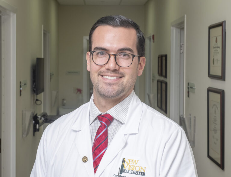
“The great thing about the cornea is it can be cleaned, and it can be fixed, unlike the retina,” said Dr. Christopher W. Seery, fellowship-trained cornea specialist at New Vision Eye Center in Vero Beach, who employs an “artistic, sophisticated surgery” technique to repair faulty corneas.
“If you think of the eye like a camera, once the film (retina) of the camera is scratched, you can’t really do anything. But the lens (cornea) can always be polished, cleaned and replaced.”
Eye infections and injuries can scar the cornea and diseases like Fuchs’ dystrophy and bullous keratopathy cause the cells in the inner layer of the cornea to quit working correctly.
If the cornea cannot be healed or repaired, an ophthalmologist may recommend a corneal transplant, in which the diseased cornea is replaced with all or part of a clear, healthy cornea from a human donor.
“Fortunately, a donated cornea doesn’t have to be cross matched for compatibility like an organ donor because the cornea is actually an avascular structure,” Dr. Seery explained. “The cornea is screened by eye banks for communicable diseases like hepatitis or HIV but not if it doesn’t have to be matched. It is also screened for quality of the cells, to make sure there’s a high density of endothelial cells and all that information about the donor cornea is sent to the surgeon a couple of days before the surgery. The eye bank screens the tissue, preps and cuts the tissue to the specific measurements supplied by the surgeon and sends it us to implant.”
According to the Cornea Research Foundation, Endothelial Keratoplasty (EK) is the preferred cornea transplant technique to restore vision when the inner cell layer of the cornea stops working properly. EK selectively replaces only the diseased layer of the cornea, leaving healthy areas intact.
There are two types of EK – Descemet’s Stripping Endothelial Keratoplasty (DSEK) and Descemet’s Membrane Endothelial Keratosplaty (DMEK). Each type removes damaged cells from an inner layer of the cornea called the Descemet’s membrane. The damaged layer is removed through a small incision and the new tissue is put in place through a small incision.
The surgeon then uses an air bubble to unfold and position the donor tissue against the patient’s cornea. The small incision is either self-sealing or may be closed with a suture or two.
The most common procedure is DSEK where the surgeon implants the back 20 percent to 30 percent of the donor cornea into the patient’s eye. Patients without other eye problems usually achieve average vision of 20/30 or better within a couple of months.
A newer form of EK, introduced in 2014, is known as DMEK. During this procedure the surgeon uses an extremely thin donor tissue, and it provides more patients with 20/20 or 20/25 than DSEK. During the procedure the patient’s existing endothelium is removed and the surgeon places the prepared donor tissue in a solution which changes it to a tinted blue color temporarily so the surgeon can better see it. The tissue is placed into a syringe type device which is inserted through the same small incision that was used for removal of the diseased tissue and the new tissue is strategically placed in the eye. Once a DMEK graft is placed into the patient’s eye, it usually curls up into a scroll which needs to be unrolled. To unroll the scroll, the surgeon uses small puffs of air and a few surgical tools to ensure the tissue is correctly placed.
“The DMEK surgery is an artistic, sophisticated surgery,” said Dr. Seery. “In order to unroll the tissue, we have to empty the fluid using a series of tapping of the surface of the cornea.
Once that is done and it’s marked in a specific way in order to make sure we can tell if it’s right side up and endothelial side down, we inject gas underneath that graft to elevate and get it to adhere to the surface of the patient’s cornea.
“The DSEK procedure uses a thicker tissue so there isn’t as much tapping and unrolling. The tissue is loaded into an injecting device and once again there is a mark that tells you which side is right up and upside down.
“As soon as the injection device is inserted into the eye, the graft is released, and it pops right open like an umbrella. Once it’s well centered, we use the same method to get it to stick to the back surface of the cornea. While it’s an easier and faster surgery, sometimes it’s worth the extra time to unroll the graft if you know it will result in higher quality vision.”
Eye surgeons decide on the best type of surgery for a patient based on the cornea’s condition and the patient’s expectations. Both procedures are done as outpatient procedures with the patient under conscious or twilight sedation. After the operation the patient is released to go home with just an eye patch.
The following day your surgeon will your check eye and give further instructions on your recovery. The stitches may or may not need to be removed and, depending on your transplant, you may have to lie on your back for a while to help the new donor tissue stay in place.
“Clinical trials are now being conducted on a new cornea transplant procedure that involves injecting the donor tissue into the eye without the positioning requirement,” Dr. Seery revealed. “We would simply inject the cells and they would uptake to the surface and get working. A single injection is all it would take, but that’s still in the future.”
Dr. Christopher W. Seery earned his medical degree and completed his internship and ophthalmology residency training at Rutgers New Jersey Medical School in Newark. To gain expertise as a cornea and refractive surgeon, he completed his fellowship in cornea, external disease and refractive surgery at Bascom Palmer Eye Institute at the University of Miami. He joined New Vision Eye Center in September 2023 and is honored to bring the latest corneal transplant procedures to Vero Beach. His office is located at New Vision Eye Center, 1055 37th Place, Vero Beach. Call 772-257-8700 to schedule an appointment.


