
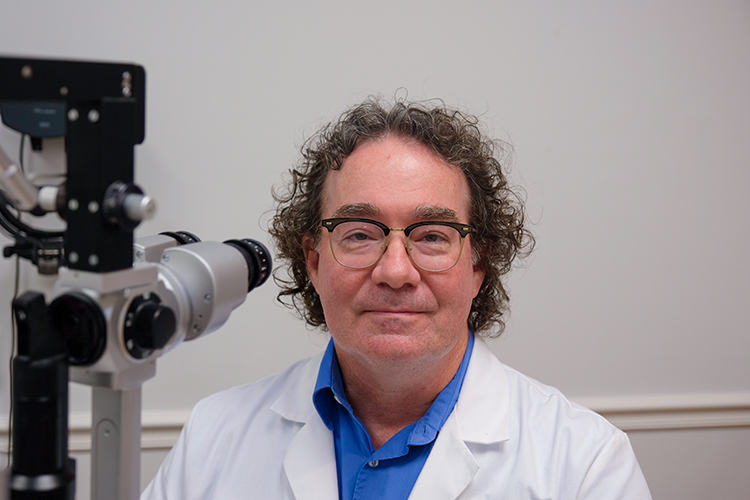
Dr. Scott Piette couldn’t be happier in his new position at the Glaucoma Institute at New Vision Eye Center. The board-certified, fellowship-trained glaucoma specialist and cataract surgeon was aware of New Vision’s stellar reputation and jumped at the opportunity to join the practice. Now he’s “having a blast” as he puts it, treating patients at New Vision’s world-class, state-of-the-art-facility.
He was attracted to ophthalmology in part because both his mother and grandmother have glaucoma and he wanted to help others like them. Glaucoma is the second leading cause of permanent blindness in the world, after blindness resulting from diabetic retinopathy. And it’s the first cause of bilateral blindness in African Americans between the ages of 55 and 75.
“Glaucoma is a progressive loss of the optic nerve tissue,” Dr. Piette said. “Think of the eye like a camera. The front of the eye is the lens that focuses the light. In the back of the eye is the retina which is like the film of the camera capturing that picture. Then it has to be taken to our computer – our brain. That is done through the optic nerve.
“The optic nerve takes all that information back to our brain, but glaucoma attacks the optic nerve like a rabbit chewing a cable and can eventually lead to blindness.”
Optic nerve damage is usually related to increased pressure in the eye due to a buildup of fluid that flows throughout the inside of the eye. This fluid normally drains out through a tissue called the trabecular meshwork at the angle where the iris and cornea meet. When the drainage system doesn’t work properly, the fluid can’t flow out at the normal rate and eye pressure increases.
“The most common type of glaucoma is primary open angle glaucoma,” Dr. Piette continued. “There are no warning signs and it isn’t associated with any symptoms until it’s pretty advanced. It slowly takes away your vision without you knowing it. Blind spots in the peripheral vision go unnoticed because the central vision remains good until the last stage of the disease when the patient goes blind.”
Damage caused by glaucoma can’t be reversed, but treatment and regular checkups can help slow or prevent vision loss. Glaucoma is treated by lowering eye pressure with the use of prescription eye drops, oral medications, laser treatment, surgery or a combination of treatments.
If eye drops and/or medications don’t bring down your eye pressure, improved drainage of the fluid within eye might be accomplished with a laser or surgical procedure. With laser therapy, the doctor uses a small laser beam to open clogged channels in the trabecular meshwork. With a surgical procedure called a trabeculectomy, the surgeon creates an opening in the white of the eye and removes part of the trabecular network.
Installing a small drainage tube the size of an eyelash is another option to drain away excess fluid to lower eye pressure. And there are other minimally-invasive glaucoma surgery options that are often combined with cataract surgery that can be discussed with your doctor.
“Acute angle closure glaucoma is another very serious condition where the natural drainage system is compromised,” Dr. Piette said. “The drain becomes closed, and the pressure will shoot up very high. This form of glaucoma does have warning signs. Your vision will suddenly get blurry, your eye will get red and the pupil will dilate. You start to see smoky vision or halos around lights. You will also feel immense pain. You need to call your eye doctor immediately because within hours you start losing vision permanently.”
According to the Glaucoma Research Foundation, Laser peripheral Iridotomy (LPI) is the top treatment for angle closure glaucoma and eyes at risk for this condition and has been used both as a treatment and prevention of the disease.
The angle is the space between the clear part of the eye (cornea) and the colored part (iris), close to their meeting point near the edge of the iris. It contains the trabecular meshwork, which normally drains fluid out of the eye. In closed angle glaucoma, the angle is closed causing increased eye pressure, which leads to optic nerve damage and vision loss. The LPI procedure creates a hole in the outer edge of the iris which opens the angle. Once the angle is widened from the procedure, the trabecular meshwork is exposed and fluid outflow is enhanced.
“You can’t avoid getting glaucoma if you are predestined to get it, so the best defense is to get a good eye exam to establish a baseline reading when you are healthy,” Dr. Piette advised. “Know your risk factors and be diligent about yearly eye exams. The earlier glaucoma is diagnosed and treated, the more sight is saved.”
The five major risk factors for glaucoma are age, family history, African-American or Latino descent, corneal thickness and elevated eye pressure.
Dr. Piette will be conducting a study for a new treatment of open angle glaucoma, working with a company called Elios on a procedure called Excimer Laser Trabeculostomy. This micro-invasive, implant-free procedure uses excimer laser light energy to create microscopic openings in the trabecular meshwork to re-establish the natural flow of eye fluid.
Dr. Piette earned his medical degree from Midwestern University, Arizona College of Osteopathic Medicine; interned at Henry Ford Hospital in Detroit; and completed his residency at Philadelphia (Pa.) College of Osteopathic Medicine and Wills Eye Hospital.
His glaucoma research fellowship was at New York Eye and Ear Infirmary, followed by additional training as a clinical fellow in glaucoma the University of Iowa Hospitals and Clinics. He can be reached the Glaucoma Institute at New Vision Eye Center, 1040 37th Place in Vero Beach, 772-257-8700.