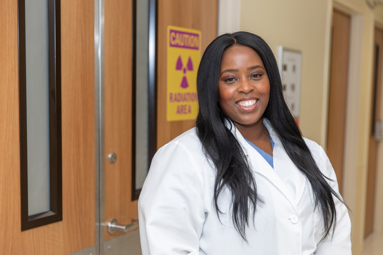
To some people, their personal image is very close to their heart.
To Dr. Mistyann-Blue Miller, an interventional cardiologist who joined Cleveland Clinic Indian River Hospital last fall, getting accurate, detailed images of other people’s hearts is far more important.
Specifically, Miller is talking about intravascular ultrasound (IVUS) and optical coherence tomography (OCT).
“Ultrasound and light energy,” Miller explains, are among the tools “we use to look into the coronary arteries.”
“We use it a lot in different settings,” Miller explains. “In cardiology, we use it to do ultrasounds of the heart, when we do the probe on the chest to look at your heart function and your valves, but we also employ it in the cardiac cath lab, which is where I do all my procedures. We do our standard angiograms and sometimes we want to take a closer look at the arteries. We can use [intravascular] ultrasound to do that.”
If you get the feeling this young Harvard-educated doctor who grew up in Miami is a big fan of this imaging techniques, you’d be right.
“IVUS gives you real-time information. In the lab, you can look at the inside of the coronary arteries to see if there’s any plaque build-up, which is atherosclerosis. You can see how much there is. You can see if there’s a lot of calcium, which is like a hard rock inside arteries, and you can determine, if someone needs a stent, how big the stent should be and how long it should be,” she said.
The American College of Radiology is just as enthusiastic about this specialized use of ultrasound technology. It says “ultrasound does not use ionizing radiation, has no known harmful effects, and can provide clear pictures of soft tissues that are not well seen on X-ray images.”
But now, as often happens in medicine, there’s something newer and possibly even better: optical coherence tomography.
OCT is an alternative imaging technology that uses a single optical fiber that emits infrared light (as opposed to the ultrasound waves in IVUS) to image the coronary artery. OCT allows for better image resolution, but this comes at the expense of tissue penetration.
Miller is fan of this technology, too. She says “it uses near infrared lights to look inside the coronary artery, and it gives us a much better resolution, much better picture” than other imaging methods. She adds that “we can’t use it with every patient,” because it requires the use of dye that some patients can’t handle.
“There are studies that show OCT gives us even better resolution than ultrasound does, [and] can help guide what we do in the cath lab and how we treat patients.”
Having access to both technologies, according to Miller, “is hard to beat.”
Miller then turns back briefly to talking stents.
Most people who have an angioplasty procedure to clear an artery also have a stent placed in the unblocked artery during the same procedure. A stent, which looks like a tiny coil of wire mesh, supports the walls of the artery and helps prevent it from re-narrowing after the angioplasty.
Like most things in modern medicine, stents have been improved since they were first used in 1986, and as Miller points out, “we use drug-eluting [stents] almost exclusively here, now, and I would venture to say that is probably true around the world.”
In a drug-eluting stent, the bare metal mesh is coated with slow-release medications that help keep the artery open. “There’s still very rare instances where bare metal stents probably make more sense, but a majority of people … [are treated] with drug-eluting stents,” Miller says.
The imaging, testing, diagnostic and procedural advances in cardiac care – some of which have taken a half a century to develop (IVUS) while others (OCT) came much faster – are a big part of what attracted Miller to the practice of interventional cardiology.
Dr. Mistyann-Blue Miller is an interventional cardiologist with Cleveland Clinic Indian River Hospital. She sees patients primarily on referral from patients’ general cardiologists. Her specialties include stenting, angioplasty and valve replacements.


