
What you can’t see can still hurt you. Worse, it can sometimes kill you.
That’s especially true when it comes to breast cancer, and efforts to detect this deadly disease go back a good deal further than most people realize.
What we now call “modern” mammography didn’t become commonly available in this country until the late 1960s, and the American Cancer Society didn’t officially endorse its use until 1976. But the very first steps in detecting breast cancer in its earliest stages actually date to 1913 when a German surgeon, Dr. Albert Salomon, published a study on using X-rays to detect “malignant entities” hidden inside breast tissue.
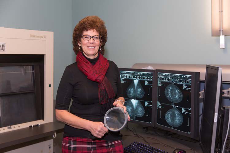
Today, Dr. Heather Nagel knows a thing or two more about detecting those malignant entities than Dr. Salomon probably ever imagined. After 23 years with Vero Radiology Associates, she has witnessed major technological leaps, including one of the most recent developments: Digital Breast Tomosynthesis, or 3-D breast scans.
“The Achilles Heel of [traditional] screening mammography,” according to the National Institutes of Health, “is the detection of cancer in women with radiographic dense breasts.
“While nearly all cancers will be apparent in fatty breasts,” NIH continues, “only half will be visible in extremely dense breasts. This results, at least in large part, from the masking or camouflaging of non-calcified cancers by the surrounding dense tissue.”
And while it may seem somewhat inappropriate to refer to a woman’s breasts as being either “fatty” or “dense,” Dr. Nagel steps in to offer a medical definition.
“It’s not a derogatory thing,” she explains. “It’s just a description of the composition of the breast. Every woman is a little bit different as far as how much glandular tissue and density she has in her breasts. When we look at a mammogram we usually quantify it, or kind of qualify it, and put them in categories.
“It’s a standard part of an imaging report,” says Nagel. “We state what their density is. In the old days we used to say mild, moderate, fatty and dense. Now we say fatty, scattered, heterogeneously dense and dense. The American College of Radiology came up with standard terminology that we all stick to it.”
Too much creativity in descriptions, Nagel warns, “can create a lot of confusion.”
Density descriptions aside, the Journal of the American Medical Association reports that 3-D mammography finds “significantly more invasive cancers than a traditional mammogram.”
Still, not everyone has embraced 3-D mammography. While Medicare approved it in 2014 and agreed to pay for it in 2015, many insurance companies still call it “not medically necessary” or “experimental,” and will not pay for 3-D scans.
Fortunately for those with stingy insurance companies, Vero Radiology charges only $64 for such a scan, which seems quite reasonable considering the group invested roughly $1 million in this new technology.
What might surprise people is that the aforementioned $1 million didn’t buy any flashy new scanners or massive pieces of equipment. It bought algorithms. Software. Software which takes the image from a 2-D mammogram and converts it, layer by layer, into a 3-D image.
Or, as Nagel explains it, “When you do 2-D, it’s like putting a book in for the mammogram. You’re seeing all the pages superimposed on one image. When we do 3-D, it allows us to look at all of those little individual pages that make up that book.”
This new 3-D technology has only been in full operation at VRA since October, but Nagel says she is more than pleased with the results so far.
“It’s basically been two months since we went live,” Nagel states, “and in that time period we’ve been collecting several cases of lesions that we might have missed on the old 2-D system. We’ve had, I think, about three cases now where the 2-D [scan] did not show the lesion, but the 3-D did.”
Nagel then adds, “Studies have shown – large studies at major academic centers where they’re looking at this – that you can find 30 to 40 percent additional invasive cancers with 3-D versus 2-D alone.”
This same technology, according to Massachusetts General Hospital in Boston, the co-developer of this particular General Electric version of 3-D mammography, can lead to earlier detection of very small, beginning-stage cancers while requiring fewer biopsies and fewer patient call-backs for additional screening.
And while Nagel freely admits that even this hi-tech software doesn’t make 3-D mammography a “perfect crystal ball,” she does say it offers much more detailed and accurate information while helping to reduce her patients’ anxiety level.
Digital Breast Tomosynthesis or 3-D breast scanning is currently available at Vero Radiology as well as in Melbourne and Rockledge through Health First Diagnostic Centers. In St. Lucie County, both Radiology Imaging Associates and the St. Lucie Medical Center offer 3-D breast scans.
Dr. Heather Nagel is the Director of the Women’s Imaging Center at Vero Radiology Associates, 3725 11th Circle. The phone number is 772-562-0163.