Over the past 50 years, accurately detecting prostate cancer through biopsies had been somewhat of a ‘hit-or-miss’ proposition.
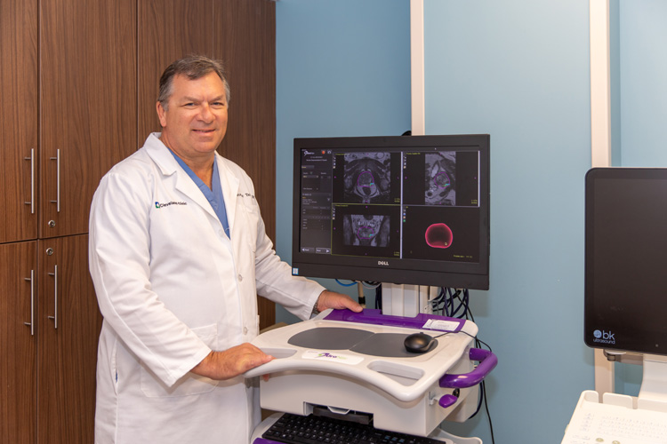
But now, according to Dr. Christopher Tardif, a urologist at Cleveland Clinic Indian River Hospital, a new hybrid technique that is technically called “multi-parametric MRI-guided biopsies” or “fusion biopsies” looks at prostate tissue three different ways to determine whether and precisely where any cancerous tissue might be hiding within the prostate gland.
A little backstory is needed here to explain how these fusion biopsies have become a game-changer in detecting and treating prostate cancer, which is the second leading cause of cancer death in men in the U.S.
From 1944 on, the prostate specific antigen (PSA) blood test – which measures levels of a protein that often goes up when prostate cancer is present – was the primary tool used to detect the disease, according to the Cleveland Clinic Health Library.
The American Society of Clinical Oncology estimates that 174,650 men in the United States will be diagnosed with prostate cancer this year and roughly 60 percent of those cases will be in men over the age of 65.
However, while those PSA tests still have some fans in medical circles, the Cleveland Clinic Library points out the test’s limitations: “Other conditions [besides prostate cancer] can elevate PSA levels. And there’s no clear-cut normal PSA level. Many men with a high PSA result don’t actually have prostate cancer, while some with low levels do.”
For collection of tissue samples after a positive PSA test, the next step was ultrasound-guided biopsies – but they, too, had their pitfalls.
“We used to do biopsies that were just ultrasound-guided,” Tradif explains, “and basically, you would put the ultrasound probe in the patient’s rectum and look around and see if you saw anything that was an abnormal-looking lesion. But most of the time we wouldn’t see anything that was an obvious pathologic finding. And so, we would usually just do systematic biopsies where you biopsy different parts of the prostate on both sides of the gland.”
“For years, the biggest frustration with the prostate biopsy was that it was blind,” says Baltimore-based healthcare system Johns Hopkins Medicine. “That is, although they were guided by ultrasound, urologists doing a biopsy really couldn’t see whether one area of the prostate looks any different from another.”
The good news? Tardif and Hopkins both say today’s new fusion biopsies are “a whole lot smarter.”
So, what are those three different ways these new MRI-guided biopsies allow physicians like Tardif to accurately target where they collect their tissue samples?
The first is the tissue signal intensity upon exposure to a strong magnet: the MRI.
The second is how well water diffuses through the tissue, and the third is how well the contrast material is taken up and how quickly it washes out of the tissue.
That’s because prostate cancer is associated with low signal intensity, low water diffusion and earlier contrast enhancement with faster washout of contrast.
“What happens is we sweep through the prostate and the ultrasound image is superimposed over the MRI image,” Tardif says. “Once we fuse those two images, we can precisely biopsy the areas that were found to be abnormal.”
The Columbia University Department of Urology adds that a radiologist will analyze the MRI images and help create a 3-dimensional image of the prostate gland, indicating abnormal areas or suspicious lesions and marking those on the images.
Those images will then be transferred electronically to the urologist’s procedure room which will allow him or her to precisely target the tissue samples to be taken.
Succinctly, Tardif says, “it allows us to see and sample areas that normally would not even have been biopsied. It’s also being used a lot to help decide if a biopsy is needed. If somebody’s got an elevated PSA level and you get an MRI and it’s completely negative, some of those people can avoid having a biopsy altogether.”
That said, Tardif also points out, “if you have a completely negative MRI, there’s still about a 10-to-15 percent chance that if you biopsy that person, they would have clinically significant prostate cancer found on the biopsy, so it’s not a perfect test, but it’s certainly a very valuable tool and really has improved the diagnostic ability of the biopsy to detect prostate cancer.”
And, as with any form of cancer, the sooner prostate cancer is detected and treated, the better the odds of complete recovery.
Medicare and most insurance will help cover the cost of these fusion biopsies.
Dr. Christopher Tardif is with the Cleveland Clinic Indian River Hospital. His office is at 3450 11th Court in the Health & Wellness building, Suite 303. The phone number is 772-794-9771.

