
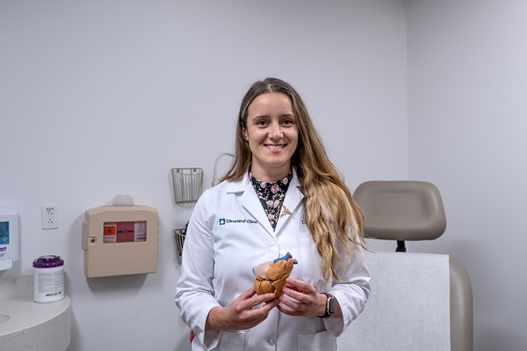
Dr. Lubka Ilieva, a cardiologist with Cleveland Clinic Indian River Hospital, is a fourth-generation physician, following in the footsteps of her great-grandfather, grandmother and mother – who is a physician at Cleveland Clinic in Ohio.
Dr. Ilieva is highly trained in utilizing the latest imaging technology to diagnose and guide the treatment of heart disease – technology her grandmother and great-grandfather could never have imagined.
One the newest technologies is transesophaeal echo (TEE), an imaging test that uses sound waves to create detailed pictures of the heart and the arteries and veins that flow to and from it.
It’s used to detect blood clots, evaluate heart valves, guide treatment for arrhythmias and many other heart conditions.
“The TEE is a higher quality imaging modality,” Dr. Ilieva explained. “The standard echocardiogram, which most people are familiar with, uses a probe on the chest wall to derive ultrasound images. Now, in order to get better visualization of certain heart structures, we can utilize new TEE technology.
“With the patient gently sedated, we pass a flexible tube into the esophagus or food pipe and into the stomach. At the tip of that tube is an ultrasound probe that delivers crisper images of certain structures of the heart. Because the esophagus is so close to the heart, we don’t have to go through the chest wall and other tissues to get to the heart and very clear images of those heart structures and valves can be obtained. We can also reconstruct 3-D images from those pictures to process a map of that structure. Basically, the TEE takes pictures inside your body, whereas the echocardiogram takes pictures from the outside.”
Doctors often use TEE when they need more detail than a standard echocardiogram can give them. The small transducer at the end of the tube produces sound waves that bounce off the different areas of the heart, making echoes. It then sends these echoes to a computer that makes them into pictures. The TEE can be combined with Doppler ultrasound and color Doppler methods to show the speed and direction of blood flow through the heart.
Cleveland Clinic offers an analogy on its website to explain the difference between the standard echocardiogram and the TEE: Imagine standing on a sidewalk and looking through a storefront window. Inside the store, near the back, you see a glass display case that contains plates with patterns, but you can’t really see the patterns because there are people in front of the case. So, you go inside and walk to the back of the store to get a better look.
While the standard echocardiogram gives your provider a storefront window view, a transesophageal echo lets your provider see directly inside the display case. The difference is between seeing a plate with a pattern and learning the pattern is sunflowers or roses.
The detailed pictures provided by TEE can help doctors see the size of the heart and how thick the walls are; how well the heart is pumping; if there is any abnormal tissue around the heart valves; if blood is leaking backward through your heart valves; or if the valves are narrowed or blocked (stenosis). It can also identify blood clots in the chambers of the heart.
“TEE is often used when there is an unexplained stroke that’s not clearly attributed to something in the brain,” Dr. Ilieva said. “The neurologist might ask us to do the procedure to see if there is any clot within the structures of the heart.
“It can also be used when someone is having heart failure due to a dysfunction in one of the valves. The TEE can give guidance on what is causing the dysfunction.
“For patients with atrial fibrillation who can’t tolerate blood thinners prescribed for stroke prevention, a medical device can be implanted in the left atrial appendage,” Dr. Ilieva continued.
“In order to size and position the device appropriately, I can do a TEE to give the operator measurements of that pocket so that the device fits perfectly. The 3-D processing and mapping can be very helpful to a surgeon. It helps him understand what his best course of action would be to fix the problem.”
Dr. Ilieva also uses another new technology called Coronary CT. It is used to diagnose coronary disease and blockage in the heart.
For years, the angiogram has been the gold standard to check for heart blockages, but it is an invasive, catheter-based procedure. For patients who may not tolerate an angiogram, a Coronary CT may be appropriate.
During this outpatient procedure, an iodine-based dye is injected intravenously and under a CT scan the physician can watch how the arteries fill during the cardiac cycle.
The dye looks bright on the scan so the doctor can see a narrowing of the passage and tell what percentage is blocked, which might be the difference between putting in a stint or treating the condition with medication.
Dr. Ilieva stressed the importance of discussing these newer, minimally invasive procedures with your cardiologist to evaluate and decide whether they are best for your specific problem.
Dr. Ilieva completed her medical education at Ohio University of Osteopathic Medicine, her residency at Swedish Covenant Hospital in Chicago and her fellowship in cardiology at Advocate Illinois Masonic Medical Center. She is accepting new patients at her office located at Cleveland Clinic Indian River Hospital’s Health and Wellness Center, 3450 11th Court, Vero Beach, and at Sebastian Primary Care, 801 Wellness Way, Sebastian. Call 772-778-8687 to schedule an appointment.